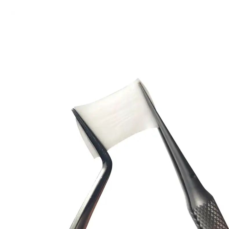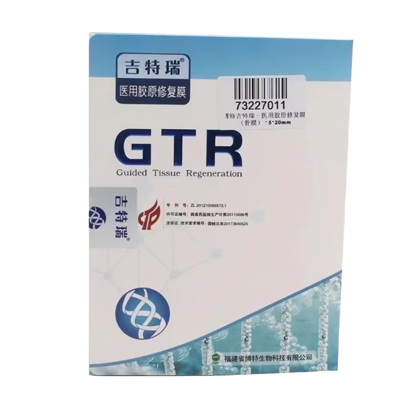
In modern restorative dentistry, a dental membrane functions as a critical physical and biological barrier that guides tissue regeneration, protects grafting materials, and allows new bone and soft tissue to form. In the oral environment, fast-migrating epithelial cells and connective tissue cells compete to populate wound sites, often outpacing slower bone-forming osteoblasts. Placing a dental membrane over a defect physically excludes unwanted cells while allowing nutrients and signaling molecules to pass through.
This biological principle, known as guided tissue regeneration (GTR) or guided bone regeneration (GBR), depends heavily on the mechanical properties and adaptation of the dental membrane. A well-fitted dental membrane creates a essential tent-like space beneath it, allowing regenerative cells to populate the void without soft tissue interference.
A dental membrane can be either resorbable or non-resorbable. Resorbable options, typically made from collagen, degrade naturally over a controlled period. Non-resorbable options, often made from polytetrafluoroethylene (ePTFE) or titanium-reinforced materials, require surgical removal. The choice of dental membrane depends on defect size, anatomy, and required healing time, but both types serve the primary goal of optimizing guided bone regeneration.
When a patient presents with insufficient bone volume at an implant site, clinicians perform bone augmentation simultaneously with implant placement. A dental membrane is placed over the grafting material to hold it in position and shield it from the surrounding soft tissue. Without a dental membrane, graft particles can migrate or resorb prematurely. For modest defects, a resorbable dental membrane is sufficient, whereas large defects require a reinforced dental membrane to prevent collapse and ensure successful guided bone regeneration.
In severe alveolar atrophy, a staged guided bone regeneration procedure is completed before implant placement. A dental membrane acts as the essential protective cover for the bone graft throughout this multi-month healing phase, resisting mechanical forces exerted by overlying soft tissues. The success of staged augmentation relies heavily on how effectively the dental membrane maintains barrier integrity during this extended regenerative period.
Periodontal disease destroys the supporting alveolar bone and attachment apparatus, creating deep intrabony defects. Surgical intervention utilizing a dental membrane encourages the regeneration of lost periodontal structures via guided tissue regeneration. By placing a dental membrane over the cleaned defect, the surgeon prevents epithelial downgrowth, allowing periodontal ligament cells and osteoblasts to repopulate the root surface.
For multi-rooted teeth with furcation defects, guided tissue regeneration using a dental membrane remains a highly predictable treatment option, particularly for Class II furcations. Additionally, in specific root coverage and recession defects, a dental membrane is used alongside or as an alternative to connective tissue grafts to maintain space, protect the healing wound, and promote stable tissue attachment.
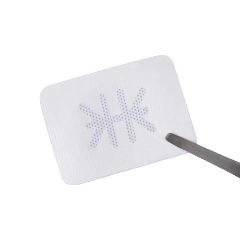
Following tooth loss, the alveolar socket undergoes rapid dimensional collapse. Alveolar ridge preservation protocols utilizing bone grafts and a dental membrane are designed to minimize bone resorption. In a standard alveolar ridge preservation procedure, the socket is filled with a bone substitute and sealed with a dental membrane. This dental membrane prevents graft migration and blocks epithelial cell infiltration, ensuring predictable alveolar ridge preservation and conserving the original bone envelope for future implant placement.
When multiple teeth are extracted simultaneously, the bone loss across the ridge can be extensive. In these high-complexity cases, a large-format dental membrane must cover a broader area, often stabilized with fixation pins. Proper adaptation of the dental membrane across multiple sites is critical, as any premature exposure of the dental membrane can jeopardize the final volume achieved during alveolar ridge preservation and full-arch augmentation.
Sinus floor elevation is performed when the posterior maxilla lacks sufficient height due to sinus pneumatization. In the lateral window approach, a clinician elevates the internal Schneiderian membrane and places a bone graft. A dental membrane is then placed over the lateral surgical window. Here, the dental membrane prevents graft particles from escaping, blocks soft tissue ingrowth, and ensures stable bone consolidation within the elevated sinus cavity.
In the transcrestal sinus floor elevation approach, a minimally invasive technique is used to lift the sinus floor. If a perforation of the Schneiderian membrane occurs during this elevation, a thin dental membrane can be placed internally to repair the tear. Incorporating a dental membrane into both lateral and crestal sinus floor elevation procedures reinforces graft containment and increases the overall biological predictability of the treatment.
Is a dental membrane always necessary when placing a bone graft? While some completely enclosed bone defects can heal without one, a dental membrane is strongly recommended for most grafting procedures. It maximizes predictability by providing essential barrier functions and maintaining space for successful guided bone regeneration.
How long does a resorbable dental membrane last in the body? The timeline varies by material. Collagen-based resorbable membranes typically degrade within 4 to 24 weeks. The degradation profile of the chosen dental membrane must match the healing rate required by the specific guided bone regeneration or guided tissue regeneration protocol.
What happens if a dental membrane becomes exposed after surgery? Early dental membrane exposure allows bacteria to colonize the surface, which can cause premature degradation or graft infection. Small exposures are managed with antimicrobial rinses, but extensive exposure may require early removal of the dental membrane, potentially compromising bone volume.
Can a dental membrane be used in combination with growth factors or biomaterials? Yes. Combining a dental membrane with biological adjuncts like platelet-rich fibrin (PRF) or growth factors enhances healing. The dental membrane contains these bio-active materials within the defect, protecting them from displacement and maximizing their regenerative potential during guided bone regeneration or alveolar ridge preservation.

 EN
EN