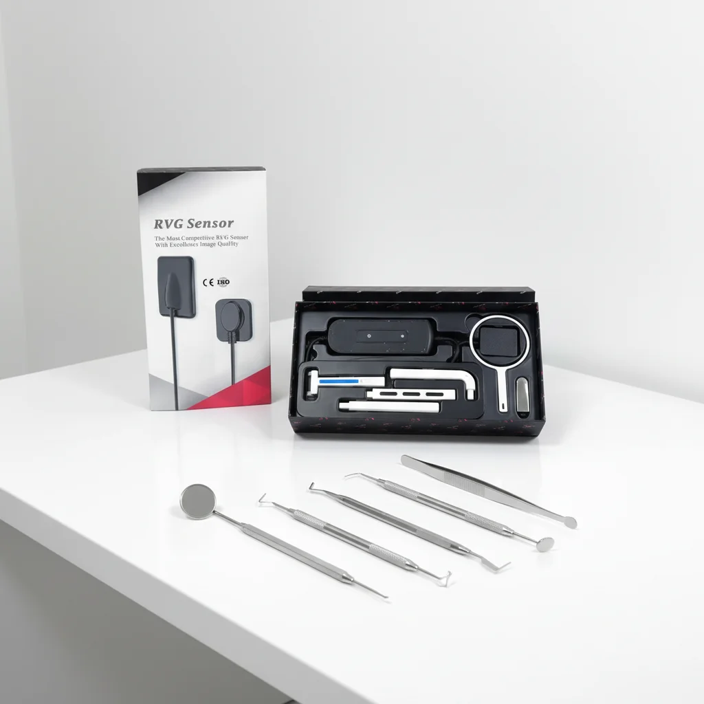
intraoral x ray
An intraoral x ray represents a fundamental diagnostic tool in modern dentistry, delivering high-resolution images of teeth and surrounding bone structures from inside the patient's mouth. This imaging technology captures detailed views that help dentists identify cavities, bone loss, infections, and other dental pathologies that may not be visible during a standard oral examination.
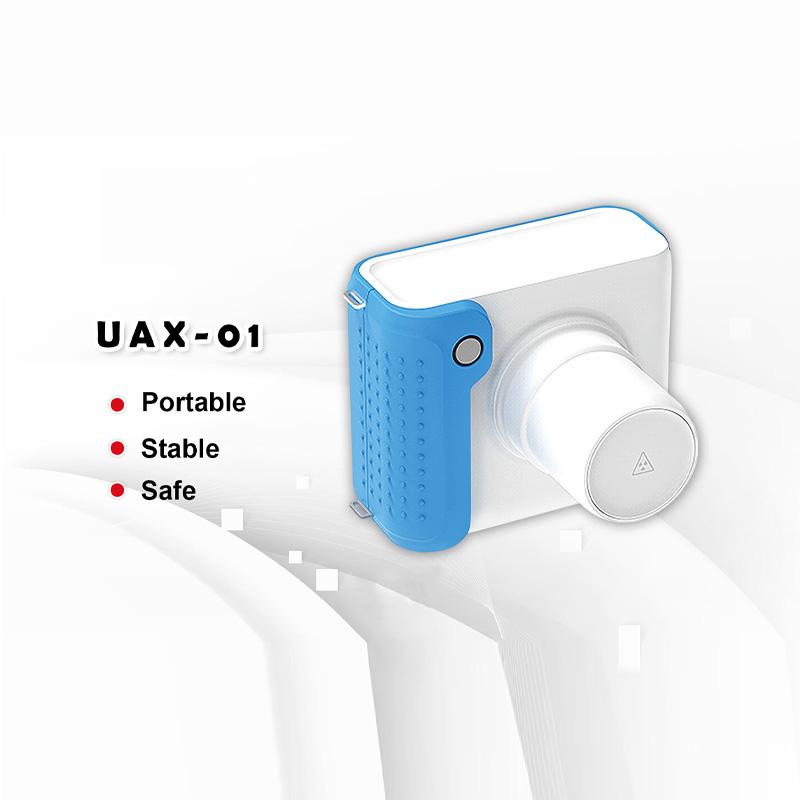
The intraoral x ray operates by positioning a small sensor or film inside the mouth, allowing the radiation source to generate precise images of specific tooth regions. Modern intraoral x ray systems utilize digital technology, reducing radiation exposure while improving image clarity and processing speed. The compact design enables practitioners to capture periapical, bitewings, and occlusal views with minimal patient discomfort.
Technological features of contemporary intraoral x ray equipment include digital sensors that provide instant image acquisition, automatic exposure controls that optimize radiation dosage, and software integration for enhanced image analysis. These systems connect seamlessly to dental practice management software, enabling efficient patient record storage and easy image comparison over time.
Applications of the intraoral x ray extend across preventive, restorative, and surgical dental procedures. Dentists rely on intraoral x ray imaging for cavity detection, endodontic treatment planning, implant assessment, and monitoring bone health. The intraoral x ray remains indispensable for comprehensive oral health evaluation and treatment documentation, making it a cornerstone of evidence-based dental practice.



 EN